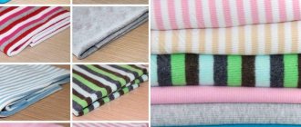
What is leather and what types of leather are there?
Leather is a natural material made from the skins of various animals.
Just as no two animals are identical, no two pieces of leather are identical. These natural features are not defects, but only add to the uniqueness of each finished product. This especially applies to exotic leather. Virtually any natural fiber other than animal and human hair can be woven into fabric, or at least thread. Also, any animal skin, except for the pseudoskin of exoskeletons in some species of fish that have a jelly-like body structure, can be turned into leather during the tanning process.
The history of mankind is the history of genuine leather products. Skin has always been a faithful assistant to man. According to archaeologists, remains of leather clothing were found in ancient Egypt, which was made back in the 13th century BC.
Leather has not lost its relevance in the modern world. With all the development of technology and the emergence of new synthetic materials, leather continues to attract more and more new supporters and admirers.
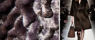
It is believed that the highest quality leather is made from the skins of cows (bull, calf). It is used for shoes, clothing, furniture upholstery, etc. Leather from sheep or goat skins (sheepskin, goat) is mainly used with hair (like fur). Leather made from pig skins is elastic, but has a persistent characteristic odor, therefore it is valued lower than others. Leather from crocodile, python, stingray, ostrich and other exotic animals is used for the production of leather goods, clothing, shoes and other business areas.
There are many classifications of leather, which differ in the type and age of the animal from which they are obtained, as well as in the methods of their processing and coloring. Here are some examples:
Saffiano is vegetable-tanned goatskin leather, lightly tanned and brightly colored.
Velor - made from leather with defects on the front surface. This is chrome-tanned leather, finished on the bakhtarma side to look like velvet using special grinding.
Suede is leather made from the skins of elk, reindeer, wild goat, etc. fat tanning. The front side of such leather is the melon side. The pile is thick, but not fluffy and without shine. The skin is soft and does not absorb water well.
Shagreen is a soft vegetable tanned leather made from the skins of sheep or goats, which has a beautiful fine relief pattern. Among the exotic leathers, this type of leather includes stingray leather.
Laika is leather made from the skins of sheep, goats, dogs, etc.
Tanning occurs with aluminum alum using salt, flour and yolk. The leather is soft, thin and is mainly used for making gloves.
Epidermis
Rice.
2. Epidermis The epidermis is a constantly exfoliating epithelial layer of the skin, which consists mainly of 2 types of cells - keratinocytes and dendritic cells. Melanocytes, Langerhans cells, Merkel cells, and intraepidermal T lymphocytes are present in small quantities in the epidermis. Structurally, the epidermis is divided into 5 layers: basal, spinous, granular, shiny and horny, differing in the position and degree of differentiation of keratinocytes, the main cell population of the epidermis (Fig. 2).
Keratinization. As keratinocytes differentiate and move from the basal layer to the stratum corneum, their keratinization (keratinization) occurs - a process that begins with the phase of keratin synthesis by keratinocytes and ends with their cellular degradation. Keratin serves as a building block for intermediate filaments. Bundles of these filaments, reaching the cytoplasmic membrane, form desmosomes necessary for the formation of strong contacts between neighboring cells. Further, as epithelial differentiation progresses, epidermal cells enter the degradation phase. The nuclei and cytoplasmic organelles are destroyed and disappear, metabolism stops, and cell apoptosis occurs when it is completely keratinized (turns into horny scales).
The basal layer of the epidermis consists of a single row of mitotically active keratinocytes, which divide on average every 24 hours and give rise to new cells in the overlying epidermal layers. They are activated only in special cases, such as when a wound occurs. Next, a new cell, a keratinocyte, is pushed into the spinous layer, where it spends up to 2 weeks, gradually approaching the granular layer. The movement of the cell to the stratum corneum takes another 14 days. Thus, the lifespan of a keratinocyte is about 28 days.
It should be noted that not all cells of the basal layer divide at the same rate as keratinocytes. Epidermal stem cells under normal conditions form a long-lived population with a slow proliferation cycle.
The spinous layer of the epidermis consists of 5-10 layers of keratinocytes, differing in shape, structure and intracellular content, which is determined by the position of the cell. Thus, closer to the basal layer, the cells have a polyhedral shape and a round nucleus, but as the cells approach the granular layer, they become larger, acquire a flatter shape, and lamellar granules appear in them, containing various hydrolytic enzymes in abundance. Cells intensively synthesize keratin filaments, which, gathering into intermediate filaments, remain unconnected on the nuclear side, but participate in the formation of multiple desmosomes on the membrane side, forming connections with neighboring cells. The presence of a large number of desmosomes gives this layer a spiny appearance, for which it received the name “spinous.”
The granular layer of the epidermis is composed of still living keratinocytes, distinguished by their flattened shape and a large number of keratohyalin granules. The latter are responsible for the synthesis and modification of proteins involved in keratinization. The granular layer is the most keratogenic layer of the epidermis. In addition to keratohyalin granules, keratinocytes of this layer contain large quantities of lysosomal granules. Their enzymes break down cellular organelles during the transition of the keratinocyte to the phase of terminal differentiation and subsequent apoptosis. The thickness of the granular layer can vary; its value, proportional to the thickness of the overlying stratum corneum, is maximum in the skin of the palms and soles of the feet.
The shiny layer of the epidermis (so named for its special shine when viewing skin preparations on a light microscope) is thin, consists of flat keratinocytes, in which the nuclei and organelles are completely destroyed. The cells are filled with eleidin, an intermediate form of keratin. It is well developed only in some parts of the body - on the palms and soles.
The stratum corneum of the epidermis is composed of corneocytes (dead, terminally differentiated keratinocytes) with a high protein content. The cells are surrounded by a waterproof lipid matrix, the components of which contain compounds necessary for exfoliation of the stratum corneum (Fig. 3). The physical and biochemical properties of cells in the stratum corneum vary depending on the position of the cell within the layer, directing the exfoliation process outward. For example, cells in the middle layers of the stratum corneum have stronger water-binding properties due to the high concentration of free amino acids in their cytoplasm.
Rice. 3. Schematic representation of the stratum corneum with the underlying granular layer of the epidermis.
Regulation of proliferation and differentiation of epidermal keratinocytes. Being a continuously renewing tissue, the epidermis contains a relatively constant number of cells and regulates all interactions and contacts between them: adhesion between keratinocytes, interaction between keratinocytes and migrating cells, adhesion with the basement membrane and underlying dermis, the process of terminal differentiation into corneocytes. The main mechanism for regulating homeostasis in the epidermis is supported by a number of signaling molecules - hormones, growth factors and cytokines. In addition, epidermal morphogenesis and differentiation are partially regulated by the underlying dermis, which plays a critical role in maintaining postnatal skin structure and function.
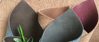
What is the difference between genuine leather, sanded and split leather?
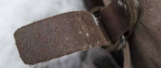
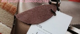
Leather with a natural grain covering - it turns out when the skin is salted, soaked (made it look like steam), until all the hairs fall out of it, then tanned and dyed. Genuine leather has almost no coating, only a little painting.
Sanded and embossed leather is made from animal skins that have natural defects. For example, if insects have bitten the skin and deposited their larvae in it, as a result of which a hole has formed in it that no longer closes, or scratches from tree branches remain on the skin, then such defects are removed by grinding during the leather production process. Then, in order for the leather to form a smooth surface, a certain coating is applied to it, the so-called primer, because of which, together with painting, the leather breathes worse and, accordingly, is less valued.
If priming is not enough to correct the leather, then an embossing process is used, in which heated leather is pressed into large slabs. By pressing you can get both a smooth leather surface and a different pattern. It is possible to visually distinguish embossed leather from non-embossed leather, but rather not by the front side, but by the mesh side.
Split is the second or third layer of leather, as if it were cut into layers. This skin may look like bakhtarma on both sides. No primer is used for its production. It is used to make sports shoes, since the foot in such shoes breathes well even under heavy loads, but such leather also gets wet and dirty.
Countries with developed chemical production know how to make very good artificial leather. But shoes made from it are more harmful than those made from genuine leather, since the foot in them breathes poorly and sweats.
Dermis
The dermis is a complex, loose connective tissue consisting of individual fibers, cells, a network of blood vessels and nerve endings, as well as epidermal outgrowths surrounding the hair follicles and sebaceous glands. The cellular elements of the dermis are represented by fibroblasts, macrophages and mast cells. Lymphocytes, leukocytes, and other cells are capable of migrating into the dermis in response to various stimuli.
The dermis, making up the main volume of the skin, performs primarily trophic and supporting functions, providing the skin with such mechanical properties as plasticity, elasticity and strength, which it needs to protect the internal organs of the body from mechanical damage. The dermis also retains water, participates in thermoregulation and contains mechanoreceptors. Finally, its interaction with the epidermis supports the normal functioning of these layers of skin.
In the dermis there is no such directed and structured process of cell differentiation as in the epidermis, however, it also exhibits a clear structural organization of elements depending on the depth of their occurrence. Both the cells and the extracellular matrix of the dermis also undergo constant renewal and remodeling.
The extracellular matrix (ECM) of the dermis, or intercellular substance, which consists of various proteins (mainly collagen, elastin), glycosaminoglycans, the most famous of which is hyaluronic acid, and proteoglycans (fibronectin, laminin, decorin, versican, fibrillin). All these substances are secreted by dermal fibroblasts. The ECM is not a random accumulation of all components, but a complexly organized network, the composition and architectonics of which determine such biomechanical properties of the skin as rigidity, extensibility and elasticity. Epidermal keratinocytes, which are closely connected to each other, are attached to the ECM proteins. They form a dense protective layer of the skin. The structure of the ECM is also capable of exerting a regulatory effect on the cells immersed in it. Regulation can be either direct or indirect. In the first case, ECM proteins and glycosaminoglycans directly interact with cell receptors and initiate specific signal transduction pathways in them. Indirect regulation is carried out through the action of cytokines and growth factors retained in the cells of the ECM network and released at a certain moment to interact with cell receptors. The ECM structural network is subject to remodeling by enzymes from the matrix metalloproteinase (MMP) family. In particular, MMP-1 and MMP-13 initiate the degradation of collagen types I and III. The density of the ECM network of the dermis is uneven - in the papillary layer it is looser, in the reticular layer it is much denser, both due to the closer arrangement of the fibers of structural proteins and due to the increase in the diameter of these fibers.
Collagen is one of the main components of the ECM of the dermis. Synthesized by fibroblasts. The process of its biosynthesis is complex and multi-stage, as a result of which the fibroblast secretes procollagen into the extracellular space, consisting of three polypeptide α-chains folded into one triple helix. Procollagen monomers are then enzymatically assembled into extended fibrillar structures of various types. In total, there are at least 15 types of collagen in the skin; in the dermis, types I, III and V of this protein are most abundant: 88, 10 and 2%, respectively. Type IV collagen is localized in the basement membrane zone, and type VII collagen, secreted by keratinocytes, plays the role of an adapter protein for attaching ECM fibrils to the basement membrane (Fig. 4). Fibers of structural collagens of types I, III and V serve as a framework to which other ECM proteins are attached, in particular collagens of types XII and XIV. It is believed that these minor collagens, as well as small proteoglycans (decorin, fibromodulin and lumican), regulate the formation of structural collagen fibers, their diameter and the density of the formed network. The interaction of oligomeric and polymeric collagen complexes with other proteins, ECM polysaccharides, various growth factors and cytokines leads to the formation of a special network with certain biological activity, stability and biophysical characteristics important for the normal functioning of the skin. In the papillary layer of the dermis, collagen fibers are located loosely and more freely, while its reticular layer contains larger strands of collagen fibers.
Rice. 4. Schematic representation of skin layers and distribution of different types of collagens.
Collagen is constantly renewed, degrading under the action of proteolytic enzymes collagenases and being replaced by newly synthesized fibers. This protein makes up 70% of the skin's dry weight. It is the collagen fibers that “withstand the blow” under mechanical influence on it.
Elastin forms another network of fibers in the dermis, giving the skin such qualities as firmness and elasticity. Compared to collagen, elastin fibers are less rigid and curl around collagen fibers. It is with elastin fibers that proteins such as fibulins and fibrillins bind, to which, in turn, latent TGF-β-binding protein (LTBP) binds. Dissociation of this complex results in the release and activation of TGF-β, the most potent of all growth factors. It controls the expression, deposition and distribution of collagens and other matrix proteins in the skin. Thus, the intact network of elastin fibers serves as a depot for TGF-β.
Hyaluronic acid (HA) is a linear polysaccharide consisting of repeating dimers of D-glucuronic acid and N-acetylglucosamine. The number of dimers in the polymer varies, which leads to the formation of HA molecules of different molecular weights and lengths - 1x10⁵-107 Da (2-25 μm), which, accordingly, have different biological effects.
HA is a highly hydrophilic substance that affects the movement and distribution of water in the dermal matrix. Thanks to this property, our skin, normally and in youth, has high turgor and resistance to mechanical pressure.
HA easily forms secondary hydrogen bonds both within one molecule and between neighboring molecules. In the first case, they ensure the formation of relatively rigid spiral structures. In the second, association with other HA molecules and nonspecific interaction with cell membranes occurs, which leads to the formation of a network of polysaccharide polymers with fibroblasts included in it. Shorter molecules of proteoglycans (versican, lumican, decorin, etc.) “sit” on a long HA molecule, like on a thread, forming aggregates of enormous sizes. Extended in all directions, they create a scaffold, contributing to the stabilization of the ECM protein network and fixing fibroblasts in a specific matrix environment. Taken together, all these properties of HA endow the matrix with certain chemical characteristics - viscosity, density of “cells” and stability. However, the ECM network is a dynamic structure that depends on the state of the organism. For example, under conditions of inflammation, HA aggregates with proteoglycans dissociate, and the formation of new aggregates between newly synthesized HA molecules (renewed every 3 days) and proteoglycans is blocked. This leads to a change in the spatial structure of the matrix: the size of its cells increases, the distribution of all fibers changes, the structure becomes looser, the cells change their shape and functional activity. All this affects the condition of the skin, leading to a decrease in its tone.
In addition to regulating water balance and stabilizing the ECM, HA plays an important regulatory role in maintaining epidermal and dermal homeostasis. HA actively regulates dynamic processes in the epidermis, including keratinocyte proliferation and differentiation, oxidative stress and inflammatory response, maintenance of the epidermal barrier and wound healing. In the dermis, HA also regulates fibroblast activity and collagen synthesis. By remodeling the matrix, HA controls the functioning of cells in the matrix, influencing their availability to various growth factors and changing their functional activity. Cell migration and the immune response in tissue depend on the action of GC. Thus, changes in the distribution, organization, molecular weight and metabolism of GCs have significant physiological consequences.
Fibroblasts are the main type of cellular elements of the dermis. It is these cells that are responsible for the production of HA, collagen, elastin, fibronectin and many other intercellular matrix proteins necessary for the formation of connective tissue. Fibroblasts in different layers of the dermis differ both morphologically and functionally. Not only the amount of collagen they synthesize depends on the depth of their location in the dermis, but also the ratio of the types of this collagen, for example types I and III, as well as the synthesis of collagenase: fibroblasts in the deeper layers of the dermis produce less of it. In general, fibroblasts are very plastic cells, capable of changing their functions and physiological response and even differentiating into another cell type depending on the stimulus received. The role of the latter can be played by signaling molecules synthesized by neighboring cells and the restructuring of the surrounding ECM.
What is pressed leather?
Pressed leather is a material produced under pressure from natural leather production waste: scraps and scraps, chrome shavings, tanning dust and other waste. Pressed leather also contains binding fibers. They can be made from any synthetic material: polyester, polyamide, polyethylene, etc. When heated, they melt and glue all the “particles” together.
Another component is synthetic thermoplastic resins. They are introduced for additional gluing and strengthening of the fibrous structure. Thanks to these resins, a material with low air and moisture permeability is obtained. The strength of such a bag made of such material is low, in contrast to a bag made of genuine leather.
Skin structure
The skin consists of 3 layers: epidermis, dermis and subcutaneous fat (SAT) (Fig. 1). The epidermis is the thinnest of them and is a stratified keratinizing epithelium. The dermis is the middle layer of the skin. Mainly composed of fibrils of the structural protein collagen. PFA contains fat cells - adipocytes. The thickness of these layers can vary significantly depending on the anatomical location.
Fig.1. Skin structure
What is the process of making leather?
The process of making leather necessarily goes through the following stages: the skin is removed, salted (if it is not salted, it will rot, since the skin is a protein (it can be boiled and eaten), so it is even transported in salted form), and then it is washed from salt and tan.
There are pores in the leather that need to be filled with a tanning agent that can stop the decomposition process and destroy bacteria. This is one of the most important processes in leather production and is called tanning.
What is tanning?! This is when leather is placed in a special drum and tanning agent is poured into it. After some time, when maintaining a certain temperature, the skin is tanned.
“Dubitel” comes from the word “oak”. Oak and willow bark are very good tanning agents. Now there are synthetic (chrome) tanning agents. Tanning substances are harmless to health.
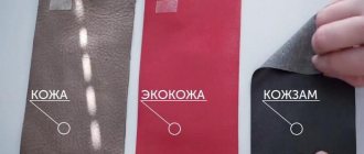
How to distinguish natural leather from artificial leather?
Thanks to the latest technologies, manufacturers have learned to make high-quality leatherette in such a way that it is almost impossible to distinguish it from genuine leather. So how else can you distinguish natural leather from artificial leather?
Leatherettes often have a textile base. Therefore, if, upon careful inspection of the seams, you see a fabric base or single protruding threads, then this is an artificial leather bag.
If you bring a lit match to natural leather, then even smoldering will not be visible on the leather, unlike leatherette, which usually begins to melt. Just be careful not to overdo this experiment - not all leather can withstand a long open fire.
There is another trick - hold the leather product in your hands: the leather will heat up, but the substitute will remain cold.
You can also identify genuine leather by its smell. He is specific and calm. If the smell is sharp and unpleasant, it means the leather goods are made from artificial leather. But it happens that even the smell of real leather is imitated due to the fact that the new generation of artificial materials includes leather crumbs.
Good skin should not have any creases. Therefore, you can bend the product (for example, the handle of a bag) with your hand and release it. If there are no creases, then this is high-quality genuine leather.
You can also drop water on your skin and see if it absorbs it. If it has absorbed and darkened, then most likely it is genuine leather; if not, it is leatherette.
Often (but not always), if the bag is indeed made from natural raw materials, then a leather sign should be stamped on the inside (or hang in the form of a tag) - an emblem that repeats the outlines of the removed skin. Honest manufacturers put a special sign on products made from artificial materials - a diamond.
And finally, the underside of real leather should be fleecy.
Stretch leather
What to look for when buying leather products?
An inexperienced buyer pays attention only to the appearance of the product, he likes it - and that’s it. But it happens that technological errors in leather processing also occur. It was poorly tanned, poorly pressed, and poorly applied primer. This product will not last long.
A discerning buyer will definitely pay attention to what the reverse side looks like - bakhtarma. From it you can immediately judge the level of leather treatment: it should be smooth, even, well and evenly dyed. And only then he looks at the face - is it natural or skillfully polished.
Be careful when choosing a leather product and it will please you for a long time.
Leather repair
Repair of leather, leather products and leather furniture, especially expensive exclusive models, should be carried out by professionals. Cheap synthetic materials commonly used for repairs, such as liquid leather, can permanently ruin your product.
In the process of using leather products, paint falls off and scuffs appear. In this case, you need to treat these places with creams of the appropriate color and then wipe them thoroughly with a brush or rag. If after such treatment the abrasions do not disappear, then it is better to seek professional help.
To prevent you from needing repairs to your favorite leather items, we recommend that you familiarize yourself with the rules for caring for leather goods and leather bags.